Click to Skip Ahead
Magnetic Resonance Imaging (MRI) is a form of advanced diagnostic imaging that generates clear 3-D pictures of the soft tissues in the body. If you want more information about your cat’s central nervous system, ears, nose and other soft tissues, MRI is ideal.
X-rays (ionizing radiation) can image bones well, provide low-detail soft tissue pictures, and are used in plain radiographs and CT scans. If your cat has a kidney or liver problem, an ultrasound can image these organs in detail. However, ultrasound cannot penetrate the bones of the spine and skull, so if detailed pictures of the brain or spinal cord are needed, MRI is the only way to go. It hasn’t been around that long, and the first vet hospitals installed them in the ‘90s.

How Does MRI Work?
An MRI machine uses the body’s natural magnetic properties to produce a picture. When your cat gets an MRI, they lie on a table and, depending on the machine, it either remains in place or slides into the MRI machine. The MRI machine then does the work of taking the 3-D pictures:
- A strong magnet is used to align hydrogen atoms in the body with the magnetic field. Hydrogen atoms are used as they are abundant in water and fat of the body.
- A radiofrequency pulse is delivered, which rotates the hydrogen atoms, disturbing their alignment with the magnetic field. The hydrogen atoms then have higher energy.
- When the radiofrequency pulse is turned off, the hydrogen atoms again line up with the magnetic field; this is called the relaxation phase. During the relaxation phase, the hydrogen atoms release the energy that they briefly gained in the form of radio-waves. The rate of the relaxation phase is characteristic of the molecule they are part of, and the frequency of the wave emitted is unique for each tiny unit of tissue in your cat’s body.
- An antenna in the machine detects these radio-waves, and the process is repeated multiple times in each section.
- The computer uses the data to estimate the chemical composition of the units of tissue and turns the data into a 3-D image that can be examined slice by slice.
In veterinary hospitals, you might find low-field or high-field MRI scanners. High-field scanners generate superior images and can be used for many views, while low-field scanners are more affordable and practical.
For low-field scanners, the cats lie on the table, and the MRI works without moving them. In high-field scanners, the cats are placed in the MRI machine, much like a human MRI.
Where Is MRI Used?
In veterinary medicine, MRI is generally only used in specialist practices. It’s the best modality to look at the brain and spinal cord tissue in cats. It helps diagnose diseases of the central nervous system. It can also help when planning radiation therapy or surgery on a tumor.
It can help determine the extent of the disease and inform treatment options. If your cat has clinical signs like seizures, poor mentation, wobbly gait, weakness, or abnormal reflexes, an MRI can help diagnose what is happening.
This modality shows the eyes and optic nerves very clearly, so problems here can be detected, too. It can also be used with CT scans for complicated joint problems, but that is more common in dogs.
- Abnormalities, including their location, size, shape, and impact on surrounding tissue, can be noted.
- A contrast material can be used to highlight the blood vessels in the brain.
- The composition of the tissues can be determined since MRI can differentiate between fluid, blood, and inflammation.
Advantages of MRI
- Unlike radiographs and CT scanners, MRI uses no ionizing radiation.
- MRI provides high-resolution images of soft tissue structures.
- MRI eliminates the effects of bones getting in the way of images.
- 3-D images are produced without needing to move the patient.
- MRI can differentiate between gray and white matter in the brain.
- MRI can differentiate different fluid types, giving more diagnostic value.
- MRI gives relatively specific diagnostic information.
Disadvantages of MRI
- Metal interferes with the image clarity in MRI.
- MRI requires highly skilled operators and interpreters, usually veterinary specialists
- MRI does not image bone well.
- Cats can have an anaphylactic reaction to contrast material used in MRI, but it is rare.
- The MRI takes a long time to complete.
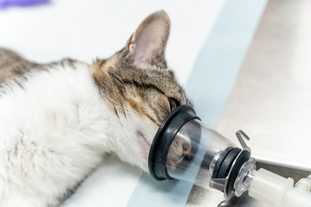
- The cat needs to be under general anesthesia to remain still for the duration of the exam.
- Special non-metal anesthesia equipment is needed, and the infrastructure needed to house an MRI machine can be very expensive. The machine itself is expensive to purchase and maintain, so the cost to owners is comparatively quite high.
- Follow-up tests like biopsies may be required once a lesion is noted.
- Cats within the tube of the high-field MRI are less accessible for anesthesia purposes.

Frequently Asked Questions (FAQ)
How Long Does a Cat MRI Take?
An MRI can take an hour or two for the imaging itself, or more in some cases, depending on the specifics of the problem. After the procedure, your cat will also need to recover from the anesthesia, which usually takes a few hours.
Do Cats Need Anesthesia for an MRI?
Yes. The MRI machine is extremely sensitive to small movements, and the cat must remain completely still for the duration of the MRI. This is only achievable under general anesthesia. The MRI scanner can also be quite noisy, which can scare cats.
What Is the Cost of an MRI for Cats?
An MRI costs a few thousand dollars. According to Lemonade Insurance, a veterinary MRI can cost between $2,500 and $6,000.

Conclusion
MRI is an advanced diagnostic imaging modality usually performed at referral and specialist veterinary practices. It can be incredibly useful for diagnosing your cat’s illness, especially if the condition is in the central nervous system. It’s essential to understand the pros and cons of MRIs before you commit to one. If the cost of an MRI is out of your budget, remember an MRI is not a cure for your cat’s disease.
Featured Image Credit: mojo cp, Shutterstock

